Digital Imaging
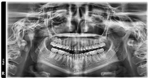
- Panoramic Image
New standard of panoramic images: Golden State X-Ray utilizes the superior VATECH PaX-i Panoramic and Cephalometric Imaging System that provides the most precise and high quality panoramic images by combining advanced digital imaging processing. This will improve the diagnostic accuracy with increased treatment planning and patient satisfaction.
Our new PaX-i next generation high resolution digital panoramic unit captures clear and sharply detailed images of the entire lower face and jaws. This digital image visualizes the mandibular condyles, maxillary sinuses, nose, maxilla and mandible. Root alignment, erupted and unerupted teeth are also displayed.
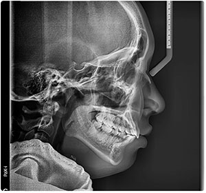 Lateral Cephalometric View
 New standard of cephalometric images: The PaX-i provides optimal images exclusively designed for orthodontics. This incredibly high resolution digital profile image shows facial relationships and measurements and can be used to predict growth patterns for orthodontic treatment planning. Unlike most typical cephalometric views, the VATECH PaX-i at Golden State X-Ray easily highlights those certain anatomical structures and points such as ANS and "A" point that are normally difficult to see clearly. The soft tissue is very well defined due to the propriety software. This view is typically useful to assist in the creation of an orthodontic treatment plan. Golden State X-Ray can create your preferred digital tracing analysis, too.
- Submentovertex View
This image shows the base of the skull, sphenoid sinuses, zygomatic abnormalities, and the angulation of the condylar heads relative to the cranial base. It is also useful in assessing symmetry of the facial structure.
- PA Skull View
The posterior/anterior image displays facial symmetry, frontal and ethmoid sinuses, occipital and facial bones, and the orbital cavities.
- Periapicals
These digital intraoral images provide a full view of the dentition, as well as detailed information about the teeth; the surrounding alveolar bone; and the tissues around the apices. They can be ordered in a full series or individually as needed.
- Occlusals
Depending on your request, we can take images of the occlusals in all three views - Maxillary Topographical, Maxillary, and Mandibular.- True occlusals will visualize the shape of the dental arches at right angles to the occlusal plane. They will display the direction of normal or displaced teeth and tooth fragments, the existence and structural influence of lesions, bone formation on the buccal and lingual surfaces of the upper and lower jaws, and the true relationship of fracture fragments. They will also expose calculi in the submandibular gland duct.
- Oblique occlusals display the area of the mandible and maxilla beyond the apex, fractures, the full extent of lesions, unerupted teeth and supernumeraries.
- Bite Wings
These can be ordered in a full-mouth series or individually as needed. Taken in either horizontal or vertical dimension, bite wings are crucial for revealing tooth decay, pulp size, improperly fitted crowns and worn restorations, and height of the alveolar crest. - Carpal Index
The wrist bone serves as a barometer for skeletal development. This image of the wrist may help determine the patient's developmental status and physical maturity for more confident treatment planning.